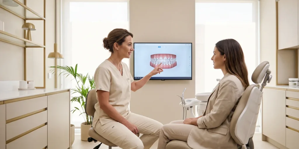
Invisalign Articles
First Invisible Orthodontic Visit in Barcelona: What's Included
What the first invisible orthodontics visit in Barcelona includes: tests, 3D scan, same-day treatment plan, budget, and what to ask before accepting it.
Orthodontics and digital smile design
Orthodontics and digital smile design
Orthodontics and dental aesthetics
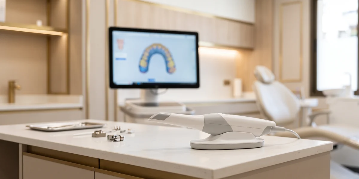
The iTero scanner is an intraoral scanner manufactured by Align Technology that generates a 3D digital model of the teeth and gums in just a few minutes, without silicone molds. Digital invisible orthodontics in Barcelona It's the tool used to take measurements, plan treatment, and display a simulation of the result. Google's artificial intelligence (AI Overviews) links it to the digital diagnosis prior to the use of clear aligners.
Beyond avoiding impression pastes, what has changed is the amount of information it provides in the same appointment: bite, wear, gum line and, in the most recent models, support to detect cavities between teeth.
Front page » blog » iTero Scanner in Barcelona: What It Is and How It Works
He iTero It is an intraoral scanner, that is, a device that photographs the inside of the mouth thousands of times per second and reconstructs a [image/image] from those images three-dimensional model exact of the dental arches, the gums and the way the teeth fit together when biting.
It is manufactured Align Technology, the same company that develops the system Invisalign and the planning software ClinCheck. This integration explains a large part of its implementation in orthodontic clinics: the scan file is sent directly to the aligner workflow, without intermediate steps or physical shipments.
The team has three parts: one head or wand that the professional passes through the mouth, a computer with the software and a screen where the model appears in real time. It does not emit ionizing radiation, unlike an X-ray, and the scan is painless.
Its use is not limited to orthodontics. It is also used for design. crowns, inlays and prostheses, implant planning, manufacturing occlusal splints for bruxism and to monitor tooth wear or gum recession by comparing scans taken separately over time.
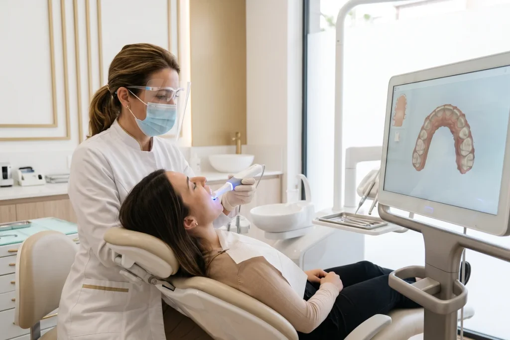
The scan is a quick procedure that requires no anesthesia or special preparation. It's helpful to understand it step by step so you arrive at your appointment with confidence.
1. Preparation. The patient sits in the chair and is asked to keep their mouth comfortably open. Sometimes a cheek retractor is used to improve visibility. Fasting and discontinuing any medication are not required.
2. Scanning of the lower arch. The professional moves the probe over the chewing surfaces, both the inner and outer parts of the teeth. The model being built appears on the screen in real time.
3. Scanning of the upper arch. The entire route is repeated.
4. Bite registration. The patient is asked to bite normally and both sides are scanned to record how the dental arches fit together.
5. Review. The software highlights incomplete areas and allows you to repeat them without starting from scratch.
The set usually includes between 5 and 10 minutes Depending on the case. You can stop at any time to swallow or rest, something impossible with a silicone tray in your mouth.
The silicone or alginate impressions For decades, they were the only way to obtain a model of the mouth. They worked, but they had limitations known to anyone who had ever used them.
Comfort. The tray filled with material occupies the entire mouth for several minutes and frequently causes nausea, especially in people with a strong gag reflex.
Precision. The material can shrink as it sets, form bubbles, detach from the mold, or deform upon removal. Then, the plaster casting introduces a second source of error. Each step adds a small deviation that ultimately affects the final fit.
Time. The physical model had to be sent by courier to the lab or manufacturer, with several days of shipping time. The digital file is sent instantly.
Repetitions. If a print job went wrong, the whole thing had to be redone. In digital printing, only the incomplete section is redone.
Conservation. Plaster models are stored, break, and deteriorate. Digital archives are stored without degradation and allow overlay scans from different dates to accurately measure how much the teeth have moved.
For the patient, the most obvious change is that the test ceases to be unpleasant and becomes part of the explanation.
Not all iTero scanners are the same, and it's important to know this when comparing clinics, because many simply advertise iTero scanners without specifying the model.
The family iTero Element, In its various versions, it has been the standard in orthodontic clinics for years. It offers color scanning, results simulation, and, in some versions, diagnostic support technology using near-infrared imaging.
He iTero Lumina is the generation presented by Align in January 2024. According to the manufacturer, it incorporates a multidirectional optic that provides a capture field three times wider than the previous model, in a a smaller 50% printhead and lighter, with faster scanning speeds and improved visualization. Restorative capabilities and gumline visualization tools have been added subsequently.
Does the model matter to the patient? In a simple case, the treatment outcome depends not on the scanner, but on the orthodontist. Where it does matter is in the comfort, in the duration of the test and in the amount of information that the professional can display on screen during the visit.
He capture field It is the mouth surface area that the scanner records with each pass. Enlarging it has practical consequences that go beyond the technical data.
Less time with your mouth open. If each pass covers more surface area, fewer passes are needed to complete the arch. In patients with limited mouth opening, joint pain, or a gag reflex, this difference is noticeable.
Better access to difficult areas. A smaller head more easily reaches the molar area and the back of the arch, which is where errors accumulate in incomplete scans.
Greater comfort for children and teenagers. A bulky wand is intrusive in small mouths, and the cooperation of the young patient directly influences the quality of the record.
More complete models. Fewer gap areas mean less software interpolation and a more reliable basis for planning moves.
It's important to keep things in perspective: a better scanner doesn't correct a misdiagnosis or poor planning. It's a tool that provides the professional with better raw data, and the final value still depends on who interprets that information and how they translate it into a treatment plan.
Scanning is no longer just about taking measurements. Integrated diagnostic support features allow for a complete oral health assessment during the same session.
Align groups these tools into the Align Oral Health Suite. These include Align X-ray Insights, an AI-assisted function for analyzing X-rays, and a visualization tool of the gum line which works by scanning to help identify and explain situations such as interproximal caries or possible gingival recession. Some models also incorporate technology of near-infrared image, designed to visualize the internal structure of the tooth without radiation.
There are two important nuances. First: these functions are of support. They indicate suspicious areas that the professional must confirm with a clinical examination and, when appropriate, with an X-ray. They do not provide a diagnosis on their own. Secondly, their availability depends on the scanner model and the software contracted by the clinic.
For the patient, the real benefit is being able to view on screen What the professional describes. Understanding why a cavity needs to be treated before starting orthodontics, by seeing it highlighted on the model, changes the conversation and makes the decision easier.
Scanning alone doesn't align anything. Its value only becomes apparent when the file enters the scheduling workflow.
Step 1. The digital model is sent along with the photographs, X-rays, and the orthodontist's prescription, which indicates the treatment objectives and clinical preferences.
Step 2. In the software ClinCheck The sequence of movements is designed: which tooth moves in each stage, by how many millimeters, and in what order. The attachments, the small composite reliefs that provide grip to the splint, and the areas of stripping where space is gained between teeth.
Step 3. The orthodontist reviews the plan, modifies it, and approves it. Automated planning tools have greatly shortened this step, even allowing the proposal to be reviewed with the patient during the same appointment.
Step 4. The aligners are manufactured according to the approved plan and delivered to the clinic.
Step 5. During treatment, control scans can be performed and overlaid on the initial scan to check if the teeth are progressing according to plan or if adjustments are needed. refinement.
The entire process starts from the same file: if the scan is incomplete, the plan carries that error from the beginning.
It's important to be clear about the boundaries, because advertising sometimes blurs them. The intraoral scanner It only records what is seen on the surfaceIt shows teeth, gums, and bite. It does not penetrate the bone or show what is happening below the gum line.
That's why It does not replace X-rays. The orthopantomography A panoramic X-ray shows the roots, supporting bone, unerupted teeth, wisdom teeth, and any lesions. Without it, tooth movement cannot be planned safely because the length and inclination of the roots and the available bone support are unknown.
The lateral teleradiograph It is necessary for cephalometric analysis in cases with bite or facial profile problems, and the CBCT, A three-dimensional X-ray is reserved for specific situations: impacted canines, bone assessment for implants, bone resorption, or joint pathology. All these tests involve radiation, so they are only indicated when they provide necessary information.
Nor does it replace a clinical examination. Tooth mobility, bleeding on probing, sensitivity, or joint pain are detected through examination, not scanning. A clinic that forgoes X-rays because it has a scanner is skipping a diagnostic step.
These are the most frequently asked questions before a scan, answered in a concrete way.
Hurts? No. The applicator head gently touches the teeth and cheeks, without any significant pressure or heat. No anesthesia is used.
Does it make you nauseous? Much less than a silicone impression, because no material is introduced and the trial can be stopped at any time. Breathing through the nose and pausing in the back of the mouth helps those with a strong gag reflex.
Is it suitable for children? Yes, and it's usually better tolerated than molds. It's common in pediatric orthodontics and in monitoring the eruption of permanent teeth.
What about with prostheses, crowns, or implants? Yes. In fact, scanning is used to design crowns and prostheses. Metal parts can reflect light and require an additional pass.
And with fixed braces in place? It can be scanned with braces, although the registration is more laborious.
Does it emit radiation? No. It uses light, not ionizing radiation, so it can be repeated as many times as necessary, even during pregnancy.
The scan generates a digital file with health data, and that has implications that are worth knowing.
That file is part of your medical record. Data protection regulations recognize the right of access and of portability, So you can request a copy. The usual format in these cases is a three-dimensional mesh file, although each system and clinic defines how and when they deliver it.
It is especially useful in two situations. The first, when ordering a second opinionWith the test results in hand, another orthodontist can assess the case without repeating X-rays. The second reason is if you change clinics mid-treatment, something more common than it seems in a city with high patient turnover like Barcelona.
There's a technical detail: treatment plans created in the manufacturer's software are linked to the clinic's professional account, so they aren't always transferable as such. Scans and imaging tests, however, do form part of your medical record.
Asking during your first visit how your data is stored, for how long, and how to request a copy is a simple check that says a lot about the clinic's transparency.
A reasonable question is whether the scanner increases the cost of treatment. In practice, scanning It is not billed separately.It is part of the initial study, which most clinics in Barcelona offer free of charge during the first visit.
What does influence the price is the type of treatment. Invisible orthodontics typically falls within a certain range. 1,800 euros in very mild cases and more than 5,000 euros In complex cases with compromised bite, depending on the number of aligners, the duration and the planned refinements.
When comparing quotes, it's important to check that they include the complete study, all the aligners, the refinements, the attachments and its withdrawal, the reviews and the retainers finals. A fixed price with everything included is usually more economical than a lower price with individual items that add up over time.
If you want to know exactly what tests and documentation are included in the initial study, it's worth reviewing What does the first invisible orthodontics visit include? before making an appointment.
Check the ranges by treatment type and the available financing options.
Between 5 and 10 minutes for both dental arches and bite registration, depending on the case and the patient's cooperation. It can be interrupted at any time to swallow or rest, and only the incomplete area can be repeated without starting over.
No. The scanner only records the surface: teeth, gums, and bite. The roots, bone, and unerupted teeth are only visible with panoramic radiography and, in specific cases, with cephalometric radiography or CBCT. Orthodontic treatment planning without radiography is not reliable.
The Lumina is the generation introduced in January 2024. According to the manufacturer, it offers a capture field three times larger in a smaller 50% printhead, with faster scanning speeds and improved visualization. For the patient, this translates primarily into greater comfort and speed.
Yes. It's much better tolerated than silicone impressions because no material is introduced into the mouth, and the trial can be stopped whenever necessary. Breathing through the nose and pausing in the molar area helps complete the impression without discomfort.
Yes. The scan is part of your medical record, and data protection regulations recognize your right to access and portability. It's especially useful for requesting a second opinion without repeating tests. The format and delivery time are determined by each clinic.
Because it summarizes clinical pages written at different times and about different models: some describe equipment from years ago, and others the current generation. The reliable way is to confirm with the clinic which specific model they use and which diagnostic functions are active.
The intraoral scanner has made the starting point of any invisible orthodontic treatment more comfortable and precise, allowing the patient to see and understand what is happening in their mouth. Even so, it remains a tool: the diagnosis, planning, and outcome depend on the orthodontist who interprets the information and the supplementary X-rays.
How useful did you find this content?
Click on a star to rate it!
Average score 5 / 5. Vote count: 1
So far, there are no votes! Be the first to rate this content.
What the first invisible orthodontics visit in Barcelona includes: tests, 3D scan, same-day treatment plan, budget, and what to ask before accepting it.

Invisalign Comprehensive in Barcelona: what complex cases it corrects, when it's not enough, how long it lasts, what unlimited aligners mean, and what the price should include.

Invisalign Lite in Barcelona: what cases it corrects, when it's not enough, how long it lasts, how many aligners it uses, and what the treatment price should include.
THE CLINIC IS CLOSED IN AUGUST.
You can schedule your first visit online now and secure your spot for a free full diagnosis in September.